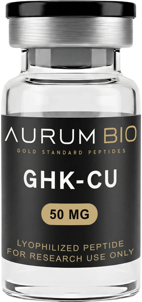

Why Is GHK-Cu Blue?
A vial of GHK-Cu is blue because copper(II) is bound inside the tripeptide's coordination environment, not because anything has been added to colour it. That fact explains the shade, but it does not make the shade a measurement. Cake density, path length and lighting move the apparent colour of an identical sample more than most people expect, and none of that touches the chemistry underneath it.
The colour is the copper, not a dye
GHK-Cu is glycyl-L-histidyl-L-lysine — a three-amino-acid peptide — complexed with a copper(II) ion. The free tripeptide has no colour of its own: like most short peptides, it has no chromophore that absorbs anywhere in the visible range, so a solution of the uncomplexed sequence looks the way water looks. Colour enters the vial only once copper(II) is coordinated by the peptide's own nitrogen atoms — the terminal amine of the glycine residue, the deprotonated amide nitrogen of the glycine–histidine bond, and the imidazole nitrogen of the histidine side chain — arranged around the copper ion in a near square-planar geometry, as solution and crystallographic studies of the complex have both shown (Freedman et al., 1982; Mehr et al., 2020). Nothing is added to produce the colour. It is a direct consequence of what copper(II) does once it occupies that specific coordination environment.
The copper coordination environment is what produces the colour wherever that complex is present. Whether the complex in a co-supplied preparation is in the same coordination state is not something the published specification addresses — and colour cannot answer it either.
What a d–d transition is, and where the absorbance sits
Copper(II) is a d⁹ ion — nine electrons occupying its outermost set of d-orbitals. In an isolated ion those orbitals sit at equal energy, but once ligands surround the metal, the electric field they create splits the d-orbitals into groups of unequal energy. A photon of visible light can move an electron between those split levels; that transfer is a d–d transition, and the tripeptide's coordination geometry is what makes it possible. For a nitrogen-coordinated copper(II) centre of this kind, that absorption sits in the orange-to-red region of the visible spectrum, measured by UV-visible spectrophotometry (Sportelli et al., 1977; Freedman et al., 1982). Absorbing there subtracts orange and red from white light passing through the solution or reflecting off the cake; what reaches the eye is the light the complex did not absorb — blue-to-violet — which is why the complex looks blue rather than orange.
That band is broad and comparatively weak next to the sharp ultraviolet absorbance the peptide bond itself produces elsewhere in the spectrum. An idealised square-planar Cu(II) complex of this kind is centrosymmetric, which makes the d–d transition Laporte-forbidden — a g-to-g transition disallowed by the relevant selection rule. It gains what intensity it has only through vibronic coupling and departures from perfect centrosymmetry, which is why its molar absorptivity sits orders of magnitude below an allowed transition and why the colour reads as comparatively muted rather than saturated. A d–d transition is a property of the coordination environment, not of the tripeptide's sequence in isolation and not of anything mixed in afterward.
Why the lyophilized cake and the solution are different shades
The compound leaves the freeze-dryer as a solid cake, not a solution, and a solid and a solution present the same chromophore to light in different ways. A dissolved sample is examined in transmission: light enters one side of the container, travels a defined path length through a reasonably uniform medium, and whatever exits the other side is what gets seen or measured. A lyophilized cake is examined in diffuse reflectance instead: it is a porous, uneven solid, and light entering it scatters repeatedly off internal surfaces before some fraction re-emerges toward the observer. Scattering competes with absorbance for every photon and does not care what colour the underlying chromophore is, so the same copper complex, at the same concentration, reads paler and less saturated as a packed powder than it does once fully dissolved. Neither shade is more correct than the other — they are two different optical situations built on the same absorbing species.
Three reasons two identical vials can look different
| Variable | What it changes | What it does not mean |
|---|---|---|
| Cake density and packing | How much internal surface light scatters against before it exits, changing how pale or saturated the cake looks under the same illumination | A difference in copper complex content between two vials of the same lot |
| Effective path length | How much coloured material sits between the light source and the eye — a taller, narrower cake reads differently from a shorter, wider one holding the same mass | A difference in what was filled into either vial |
| Light source | The colour temperature of the ambient light shifts the hue perceived from any coloured object, independent of that object's own absorbance spectrum | A change in the vial's chemistry between one look and the next |
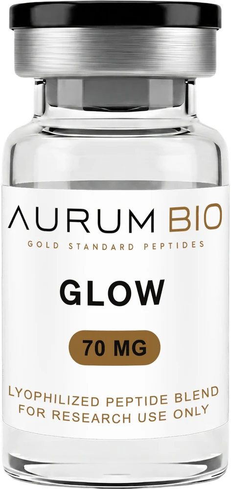
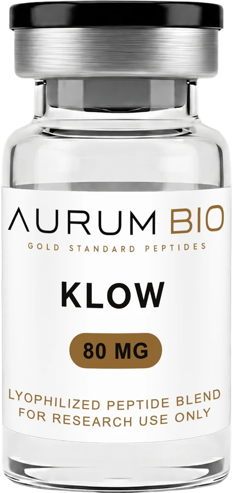
The table above holds the complex itself constant in every row — only the light's path or the light's source changes. There is a fourth cause that does not belong in it, because it changes the complex rather than the light: Aurum's own handling notes for GHK-Cu, GLOW and KLOW state, verbatim, "Protect from light — copper complexes are photosensitive." Where the rows above change how light travels to or from an unchanged sample, photolytic exposure changes the sample itself — a chemical event, not an optical one — which is the reason the handling instructions call for protection from light in the first place.
The failure mode: grading a vial by its shade
The failure mode this invites is judging material by its shade — reading colour as a stand-in for a quality decision it was never built to support. Apparent colour in a lyophilized cake is a function of cake density, packing and path length before it is a function of anything chemical, which is exactly why two vials drawn from the same lot can read differently side by side under identical light without either one being wrong. Colour is a property of the copper coordination environment, and that environment can persist through changes an eye has no way to register.
That scope matters because a separate note in this journal, on broken and discoloured lyophilized cakes, states that yellow, brown or patchy discoloration is one of the few visual observations worth raising with a supplier. Read next to each other, the two statements look like they disagree. They do not: they apply to different baselines. Where a solid is nominally white, any visible hue is new information — presence against a baseline of absence, which is why yellow, brown or patchy discoloration is worth raising. Where a solid is a coloured coordination complex, the baseline is already a strong visible absorbance, and differences in depth of shade are dominated by cake density, path length and illumination rather than by a change in what the material is (Patel et al., 2017). One rule is about presence; the other is about degree.
GHK-Cu is not exempt from the first rule. A patchy, brown or green-brown appearance in a copper complex is still a discontinuity worth raising — that is a change in hue, not a difference in the depth of an already-expected blue, and the note above does not defend it. It defends a vial that reads a shade paler or deeper than another vial of the same lot. It says nothing about a vial that has turned a different colour altogether.
What an identity test does that an eye cannot
An identity determination answers a different question than an eye does, and it answers it independent of how the vial looks. It does not read absorbance in the visible band at all — it asks whether the molecule present matches the molecule intended, using a method built for that question rather than adapted from one designed to do something else. What that kind of determination actually requires, and what it can and cannot rule out, is covered in the note on HPLC and mass spectrometry elsewhere in this journal. The point that matters here is narrower: that method returns the same result whether the cake in front of it happens to look pale or deep blue, because colour was never part of what it measures.
What this does not establish
Colour, on its own, cannot establish concentration — a paler solution might be more dilute, or it might be the same concentration examined through a shorter path length, and telling those apart requires a calibrated measurement rather than a comparison by eye. It cannot substitute for an identity or purity determination, for the reason above. It cannot detect a change that leaves the copper coordination environment and the complex's concentration untouched, and plenty of ways a sample could be compromised do exactly that without shifting the visible spectrum at all. And it cannot be compared meaningfully between two vials, two lots or two suppliers unless concentration, cake structure and lighting are all held constant — a condition that holds inside a spectrophotometer and almost nowhere else.
What Aurum specifies, and what it does not
Aurum's published specification for GHK-Cu carries the same generic assurance stated across the catalogue — independently assayed for purity and identity — with no method named and no distinction drawn from peptides that carry no metal centre at all. A peptide-level determination of that kind would not distinguish GHK-Cu from decoppered GHK: the copper is the thing that makes the complex blue, and it is not the thing a peptide-focused identity and purity assay is built to see. There is no stated hue, no shade range and no visual acceptance criterion anywhere in the published material — Aurum does not release or reject material on colour. The product page's description of a distinctive deep blue colour in solution is descriptive language about what the complex looks like once dissolved, not a specification a vial can be checked against. No visible-spectrum data and no lot-level copper coordination state are published.
References
- 01Freedman JH, Pickart L, Weinstein B, Mims WB, Peisach J Structure of the Glycyl-L-histidyl-L-lysine–copper(II) complex in solution. Biochemistry, 1982.
- 02Mehr A, Henneberg F, Chari A, Görlich D, Huyton T The copper(II)-binding tripeptide GHK, a valuable crystallization and phasing tag for macromolecular crystallography. Acta Crystallographica Section D: Structural Biology, 2020.
- 03Sportelli L, Neubacher H, Lohmann W On the influence of aromatic residues on the interaction of copper (II) with small peptides containing aromatic amino acids: ESR and optical studies. Zeitschrift für Naturforschung C, 1977.
- 04Patel SM, Nail SL, Pikal MJ, Geidobler R, Winter G, Hawe A, Davagnino J, Rambhatla Gupta S Lyophilized Drug Product Cake Appearance: What Is Acceptable?. Journal of Pharmaceutical Sciences, 2017.
Every citation links out to the paper on PubMed. Identifiers are omitted deliberately rather than reproduced from memory, so where we do not hold a verified PMID or DOI the link is a PubMed search for that exact title — it resolves to the paper without anything being invented.
FOR RESEARCH USE ONLY · NOT INTENDED FOR HUMAN CONSUMPTION. This article describes compounds and the research literature in which they appear. Nothing here is a recommendation, protocol, or statement of effect.